Beyond Insulation:
how myelin orchestrates the life of an axon.
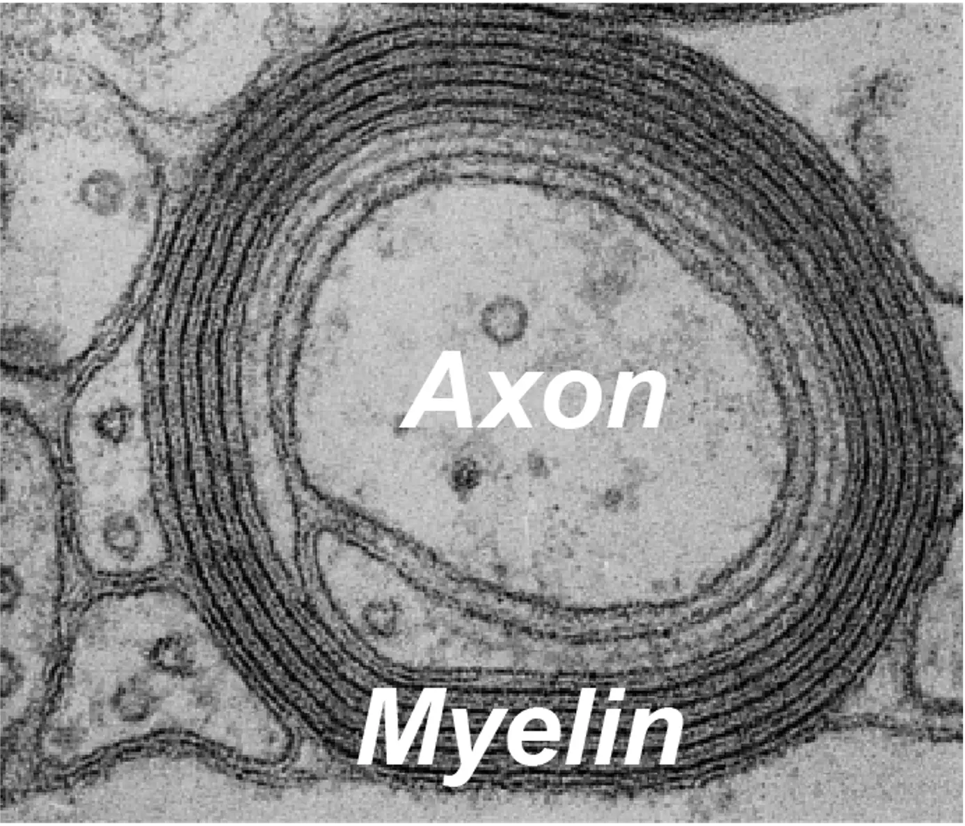
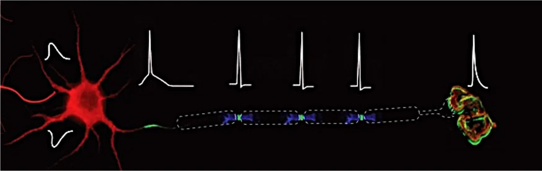
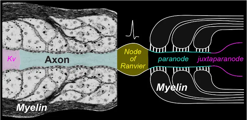
Myelination is perhaps one of evolution’s greatest inventions in the vertebrate nervous system. By enabling rapid, energy-efficient transmission of action potentials through saltatory conduction, myelin allows remarkable signaling speeds without needing the gigantic axons found in animals like squid.
In the central nervous system, oligodendrocytes (OLs) are the sole myelin-producing cells. Abnormalities in myelin and OL function are implicated not only in demyelinating diseases such as multiple sclerosis, but also in neurodegenerative and psychiatric disorders.
Remarkably, a single OL can myelinate up to 60 distinct axons, establishing intimate connections with multiple neurons simultaneously. OLs are far more than passive insulators of axons. Through these interactions, OLs contribute to nervous system development, homeostasis, plasticity, and repair. Despite their fundamental importance, how neurons and glia communicate at the molecular level remains poorly understood.
Some of these questions keep us awake at night...
Could a single OL integrate information from multiple axons? Are these connections merely structural anchors, or do they serve as active hubs for intercellular communication? How might these interactions shape nervous system plasticity, resilience, and vulnerability to disease?
The paranodal paradox
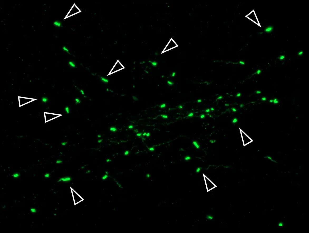
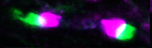
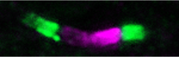
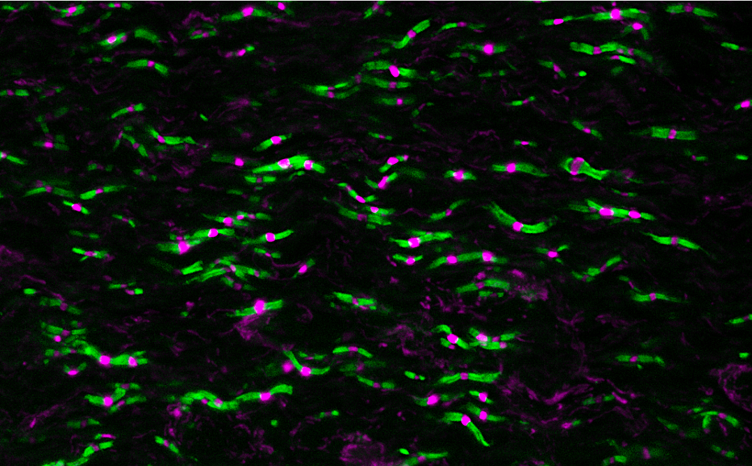
To move or not to move, that is the question. During development, neighboring myelin sheaths elongate toward one another, bringing heminodal protein clusters together to form mature nodes of Ranvier. Once established, these specialized axon–glia interfaces can remain remarkably stable throughout life, even as the nervous system adapts to changing physiological demands and experiences. How do neurons and glia reconcile the seemingly opposing demands of structural plasticity and lifelong stability? We seek to uncover the molecular mechanisms that allow these connections to remodel when necessary while preserving their structural and functional integrity.
A second chance
Despite their remarkable stability, neuron–glia interfaces can become vulnerable to aging, injury, and diseases such as multiple sclerosis. Is this merely a consequence of pathology, or could it be an early driver of neurological dysfunction? Can we repurpose developmental mechanisms to rebuild these connections in adulthood? Axonal regeneration or remyelination alone may not be enough to fully restore function. Using CNS injury and disease models, we seek to understand the mechanisms underlying their vulnerability and resilience, with the ultimate goal of repair. A second chance for neurons and glia to reconnect may one day offer patients a second chance at functional recovery.
Pushing the boundaries
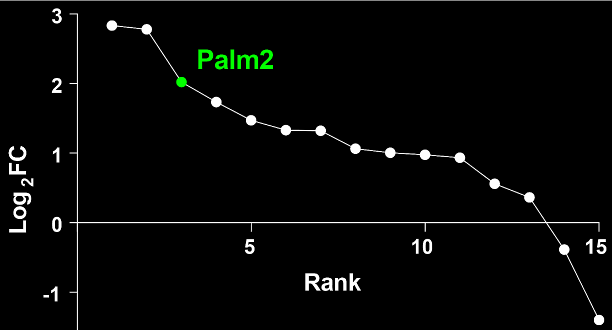
In the nervous system, molecular function depends not only on what molecules are present, but also on where, when, and with whom they interact. Conventional approaches often lack the spatial and temporal resolution needed to capture these interactions in their native physiological context. We therefore aim to develop innovative technologies to enable subcellularly resolved molecular discovery and manipulation in vivo, integrating endogenous proximity proteomics, AAV-mediated genome editing, RNA manipulation, and lipid profiling. By bridging unbiased molecular discovery with rigorous mechanistic validation in vivo, we aim to push the boundaries of what is experimentally tractable.